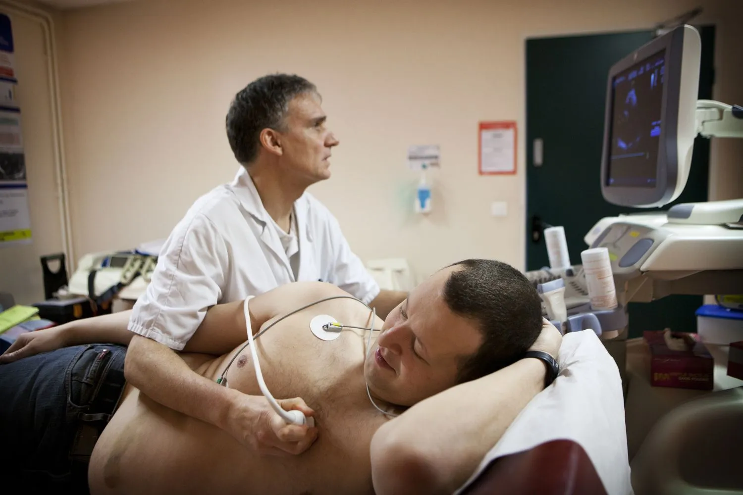
2D ECHO Service available at Silver Birch Diagnostic Center
A 2D echocardiogram, often abbreviated as 2D echo, is a medical imaging technique that uses ultrasound waves to create a two-dimensional cross-sectional moving image of the heart. It provides detailed visual information about the structure and function of the heart in real-time. The term "echo" refers to the use of sound waves to generate images, and "2D" signifies the two-dimensional nature of the images produced.
During a 2D echo, a transducer (a handheld device that emits and receives ultrasound waves) is placed on the chest or sometimes inside the esophagus. The ultrasound waves bounce off the heart structures, creating echoes that are then processed to generate detailed images of the heart's chambers, valves, and surrounding tissues. This imaging modality is commonly used in cardiology to assess cardiac anatomy, detect abnormalities, evaluate heart function, and diagnose various cardiovascular conditions such as heart valve disorders, congenital heart defects, and heart muscle abnormalities.

What Our Patients Say


One of Good diagnostic centre in our area.. Staff are pretty Good and Polite. Quick service and quality Diagnosis...silwer birch Diagnosis nawle bridge


Best service all staff are good and excellent service


Excellent staff and excellent service in affordable prices than other centers ..everyone should visit to this ..


One of Good diagnostic center in our area.. Staff are pretty Good and Polite. Quick service and quality Diagnosis...


Best diagnostic center in Pune . All staffs are very polite and supportive. I recommend to my friends and family.


One of the Best diagnostic centre at Narhe Ambegaon area. Services that I received are excellent. Medical staff, technicians are very supportive. Centre premises maintained very clean, good environmen...
